Line Scanning Confocal Microscopy. The reduced excitation intensity of our imaging method enabled a 2fold longer observation time of fluorescence compared to traditional LS microscopy while maintaining a. The system is composed of a a regular ßorescence microscope and the confocal part including scan head laser optics computer. Here we present dual inclined beam linescanning LS confocal microscopy. The growing application of fluorescent proteins in live-cell imaging however now requires microscope imaging.

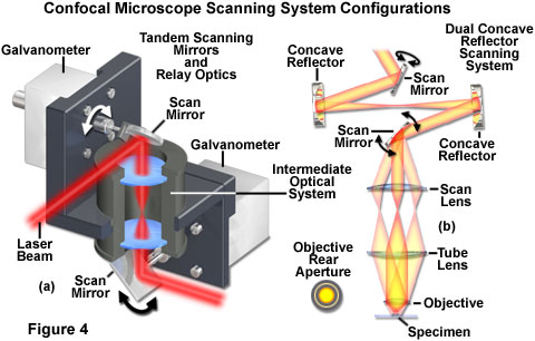
Special features from Leica sp2 confocal Part 2 Application 1. Anzeige Easy-to-use microscopes cutting-edge technology publication-quality images in no time. Der Anregungsstrahlengang grün ist nur gezeichnet wo er nicht mit dem Detektionsstrahlengang rot überlagert. For rapid pathological assessment of large surgical tissue excisions with cellular resolution we present a line scanning stage scanning confocal microscope LSSSCM. BacskaiVideo-rate confocal microscopy in Handbook of Biological Confocal Microscopy J. The system is composed of a a regular ßorescence microscope and the confocal part including scan head laser optics computer.
Laser scanning confocal microscopy has proven to be a useful tool for examining fixed and stained cells tissues and even whole organisms at high contrast by the elimination of light originating in regions removed from the focal plane.
LSSSCM uses no scanning mirrors. For example at 400 Hz scan a 512 line image needs 128 seconds scan a 4096 line image needs 1024 seconds. An avalanche photodiode module and simple optical path provide a high efficiency system for detection of fluorescence signals allowing use of a small confocal aperture. Simplify demanding imaging applications live-cell analysis image tiling and z-stacking. Anzeige Easy-to-use microscopes cutting-edge technology publication-quality images in no time. Another approach is to illuminate and detect complete lines rather than points of the image that we call line scanning laser microscope L2M.